

mNSET Device for Mice 60010 Resource Page

IMPORTANT! Before beginning the mNSET 60010 device technique, please read carefully the: mNSET 60010 Technical Support Letter, mNSET 60010 Instructions, mNSET 60010 Helpful Hints, and mNSET 60010 FAQs.
• mNSET 60010 Technical Support Letter [PDF only]
• mNSET 60010 Instructions [PDF]
• mNSET 60010 Helpful Hints [PDF]
• mNSET 60010 Frequently Asked Questions [PDF]
• mNSET 60010 Publications [PDF]
• mNSET 60010 Presentations
• mNSET 60010 Videos [Complete Demonstration] & [Quick Procedure Only]
• mNSET 60010 Webinar
The mNSET™ Device 60010 is manufactured in the USA, solely for ParaTechs Corporation, and is EtO (Ethylene Oxide) sterilization processed. Patent Information: Non-Surgical Embryo Transfer Method and Apparatus, United States Patent 9,615,903. [PDF]
International shipments are subject to customs duties and taxes. The Recipient (Importer) of such goods is responsible for paying all related charges. Please read our Goods Customs Duties and Taxes Statement. [PDF]
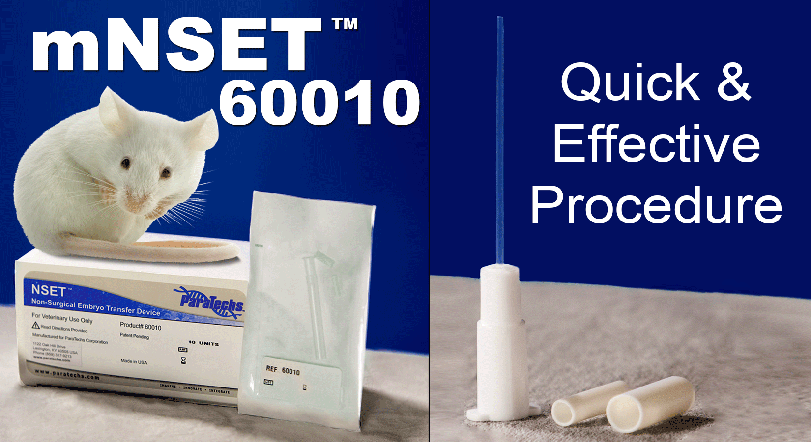
mNSET Device for Mice 60010 Instructions for Non-Surgical Embryo Transfer
Non-Surgical Embryo & Sperm Transfer Device for Mice
Product Information
Catalog #: 60010
Unit Quantity: 10 devices per box (sold in box quantities only) 1 Box = 1 Unit
Patent Information: Non-Surgical Embryo Transfer Method and Apparatus, United States Patent 9,615,903. [PDF]
FOR TECHNICAL SUPPORT
Please Call: +1(859)317-9213 or Email: info@paratechs.com
Intended Use
This device is used for non-surgical transcervical transfer of mouse embryos into female recipient mice. For research purposes only.
Not intended for human or animal diagnostic or therapeutic uses.
Other Uses: The mNSET device (60010) can also be used for artificial insemination or other material/pathogen transfer into recipient female mice. However, the mC&I device (60020) is recommended for transfer of liquids over 10µl.
Handling
Devices are single use only. Discard after use.
Reagents (not included)
M2 media (Millipore Cat #MR-015-D) equilibrated to 37˚C.
Prior to mNSET transfer
For standard use, mouse embryos should be at blastocyst stage on the day of mNSET transfer; recipient female mice should be 2.5 days post coitum (dpc). Pseudopregnant females are produced by mating with vasectomized males or by cervical manipulation as described by Stone and Srodulski (2023)1.
Embryo Transfer Procedure
1. Place a 20μl drop of M2 onto the lid of a tissue culture dish.
2. Under a microscope using reflective lighting, load 10–20 blastocysts into the media drop using a standard embryo handling pipet (90010). (Note: The optimal number of embryos to transfer will vary depending upon the donor mouse strain and embryo health/ manipulations.)
3. Secure the mNSET device onto a P-2 pipette that has been set to 1.8μl. Remove the protective catheter cover. Important: The mNSET hub must be firmly seated on the P2 pipette to effectively uptake and dispense liquid. Please be sure to firmly attach the device using pressure and a slight twisting motion. This will ensure proper placement.
4. Press the pipette plunger to the first stop, lower the catheter tip into medium and slowly pull the embryos into the tip of the mNSET device. Remove the mNSET tip from the medium.
5. Carefully set the volume to 2.0μl to create a small air bubble at the mNSET tip. (This helps prevent embryos from wicking out during mNSET insertion into mouse.) Avoid touching the mNSET tip to any surface prior to embryo transfer.
6. Place the recipient female on the top of a cage, allowing the mouse to “grab” the cage bar surface. Grasp near the base of the tail using your thumb and forefinger and angle the tail upward while stabilizing the animal on both sides as shown below.

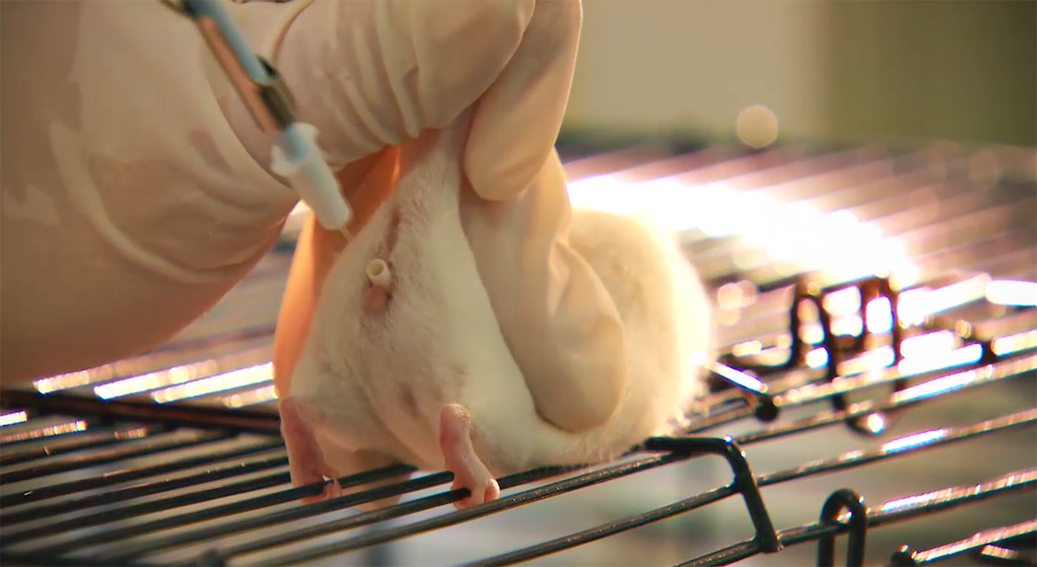
7. Gently place the small speculum into the vagina.
8. Optional: Remove small speculum and replace with the large speculum. If desired, use an adequate light source and visualize the cervix.
9. While holding the female mouse with one hand as described in step #6, carefully pick up the pipette and insert the mNSET tip into the speculum and through the cervix. Once the mNSET device hub contacts the speculum, expel embryos by pressing the plunger to the first stop. Do not release the plunger.
10. Gently and slowly remove the mNSET device and then remove the speculum. Return the mouse to its cage. No post-procedure monitoring is required.
References
1Stone BJ, Srodulski SJ. Inducing Pseudopregnancy in Female Mice Without the Need for Vasectomized Males Prior to Non-Surgical Embryo Transfer or Artificial Insemination. J Vis Exp. 2023 Jul 7; (197):10.3791/65477. doi: 10.3791/65477. PMID: 37486140; PMCID: PMC10642351.
This product is intended for research purposes only.
CAUTION: Not intended for human or animal diagnostic or therapeutic uses.
Purchase does not include or carry any right to resell or transfer this product either as a stand-alone product or as a component of another product.
PARATECHS COROPORATION LIMITED WARRANTY
ParaTechs warrants that, at the time of shipment, the Product will conform to the specifications that accompany the Product. This warranty limits ParaTechs’ liability to replacement of the Product.
PARATECHS MAKES NO OTHER WARRANTIES, EXPRESS OR IMPLIED, WITH RESPECT TO THE PRODUCT; INCLUDING ANY WARRANTIES OF MERCHANTABILITY OR FITNESS FOR ANY PARTICULAR PURPOSE OR THAT THE PRODUCT DOES NOT INFRINGE ANY PROPRIETARY RIGHTS OF ANY THIRD PARTY.
For further information on other ParaTechs products contact us at info@paratechs.com or call +1(859) 317-9213.
United States Patent 9,615,903 Revised: April2026
mNSET Device for Mice 60010 Helpful Hints
The following are hints and suggestions from our scientists, technicians and customers which have proved helpful.
1. Read all “Helpful Hints” and the mNSET for Mice 60010 instruction insert carefully before beginning your mNSET trials. The instruction insert can be found in each box of NSET.
2. Prior to actual experiments, practice the mNSET technique on 2.5 dpc pseudopregnant mice without embryos.
3. mNSET is designed to fit snugly on a Rainin Classic PR2, 0.1-2µl or Gilson Pipetman P2, 0.2-2µl pipette for loading embryos and precise measurement of media into the tip of the device.
4. We suggest using conscious, calm, and unagitated mice. This makes it easier to get the mouse in a natural position to find and enter the cervix. The female mouse in the video on our website is not sedated.
5. We use CD1 mice and highly recommend using this strain for your pseudopregnant recipients. We suggest using mice that weigh ≥26g and are at least 60 days old.
6. Embryos should be incubated to blastocyst stage (e3.5) since the device transfers them to one of the uterine horns and not the oviduct. Some end users have been successful using morula embryos.
7. Select a media you have used which gives you the best success in incubating your embryos. For example, use M2 or KSOM medium and transfer 12 to 20 embryos.
8. The mNSET device is only able to pass the cervix during certain phases of estrus and at 2.5 dpc in pseudopregnant mice. Therefore, we strongly recommend the use of 2.5 dpc pseudopregnant mice (after the plug has fallen out) for training purposes and embryo transfers using the mNSET device.
9. We find it relatively easy to keep the female still and reduce squirming by placing the mouse on top of the cage with a wire rack so she can grab the cage bar surface. Use the holding technique as described in #6 of the mNSET instructions and also demonstrated in the mNSET videos found on our webpage: https://paratechs.com/collections/art-devices/products/nset-device-formice#mnset-60010-videos.
10. We do not recommend the use of lubricants. You may use sterile water or culture media to moisten the specula then shake off excess before insertion into the vagina.
11. Gently insert small speculum into the vagina. The mouse will innately push the speculum out a little. Gently press it back in place so the mNSET device can pass through the cervix and into one of the uterine horns.
12. Optional: Remove small speculum and replace with large speculum. If desired, use an adequate light source such as a gooseneck light and visualize the cervix.
13. Be patient and do not apply too much pressure when finding and penetrating the cervix with themNSET tip. This could cause tissue damage and will likely bend the mNSET device tip making it nearly impossible to use. If the first attempt to insert the mNSET is not in the correct location, gently reposition the device and repeat.
14. Embryo loss may occur if tip gets bent due to too much pressure applied while finding the cervical opening. Again, gentle repeated attempts are pertinent to mNSET success.
15. You will know the device is properly inserted through the cervix into the uterus when the hub of the mNSET device touches the end of the speculum.
16. To expel your embryos, press the pipette plunger to the first stop. Count to 3. Do not release plunger.
17. Slowly remove mNSET without releasing pipette plunger. If plunger is released prior to removal, some embryos could be pulled back into the tip.
18. Inspection of the mNSET tip under a microscope after use is good practice. The clear tip mNSETallows visualization inside the tip.
The device is designed for a one-time use only. Repeated use will clog the mNSET tip with cervical tissue. Reuse may render the catheter pliable and no longer rigid enough to pass the cervix. Thus, potentially depositing embryos in the vagina and not the uterine horn as intended.
Contact us with any questions by phone or email info@paratechs.com. We are always happy to help.
mNSET Device for Mice 60010 Frequently Asked Questions
1. Can I reuse the mNSET device to transfer embryos into multiple mice?
ParaTechs does not recommend using the mNSET device for more than one transfer. Tissue from the mouse reproductive tract tends to clog the catheter. Reuse also renders the catheter pliable and no longer rigid enough to pass the cervix, thus depositing embryos in the vagina. When the device is used multiple times there may be a noticeable drop in the success rate of embryo transfer.
2. What mouse strain should I use as my embryo recipients?
ParaTechs recommends CD1 or ICR mice. For best results, the recipients should weigh ≥ 26g and be over 60 days old.
3. What day post coitum (dpc) should the recipient mice be?
We strongly recommend the use of 2.5 dpc pseudopregnant mice for both training purposes and embryo transfers using the mNSET device. It is possible for the mNSET device to pass through the cervix of a pseudopregnant mouse 1.5 dpc. However, the success rate of embryo transfer at that time has shown to be lower than using the recommended 2.5 dpc pseudopregnant female.
4. What developmental stage should my embryos be for transfer?
Embryos should be in a later developmental stage than the reproductive tract of the pseudopregnant female. For instance, blastocysts (e3.5 days after fertilization) should be transferred into a 2.5 dpc pseudopregnant recipient.
5. Is anesthesia required to perform the mNSET procedure?
No. ParaTechs does not recommend the use of anesthesia. A calm conscious animal can be positioned so that the mNSET device catheter can easily pass the cervix. The mouse in the demonstration video on our website is not anesthetized: https://paratechs.com/collections/art-devices/products/nset-device-for-mice#mnset-60010-videos. Using an unanesthetized mouse also makes the procedure faster and easier while eliminating the risks and stress of anesthesia.¹ Anesthesia may be helpful for training purposes but need not be used under ordinary conditions.
6. Should I use a lubricant during the mNSET procedure?
No. Lubricants can clog the mNSET catheter and prevent the correct placement of embryos in the uterine horn. The specula maybe moistened with sterile water or culture media prior to insertion, but even this is unnecessary. If moistening the specula, be sure to shake off any excess moisture before inserting the devices into the vagina.
7. I’m having trouble locating and passing through the cervix. What should I do?
Be sure that your recipient female is 2.5 dpc pseudopregnant. Use gooseneck lighting to inspect and locate the cervical opening before inserting the mNSET catheter, as this will help you position the device correctly. It may be helpful to practice passing through the cervix of 2.5 dpc pseudopregnant females before attempting embryo transfer.
8. How many embryos should I transfer into each recipient mouse?
For most transfers, ParaTechs recommends transferring 12-20 embryos to each recipient mouse. (Note: optimal number of embryos to transfer will vary depending upon mouse strain and manipulations embryos have received.)
9. I performed a non-surgical embryo transfer, but when I removed the mNSET device, the catheter was bent. What happened?
A bent catheter likely means that the catheter did not enter the cervix or that you applied too much pressure while trying to locate the cervical opening. If the catheter is bent, it is unlikely that the embryos were deposited into the uterine horn of the mouse. It is important to use gentle pressure when locating the cervical opening.
10. Can I use mice multiple times as embryo recipients?
Studies by ParaTechs have shown that it is possible to perform multiple mNSET procedures on a female recipient and obtain up to three litters. However, there was a decrease in pregnancy rate and embryo transfer efficiency after the first litter.
11. Can the mNSET device be used for Artificial Insemination (AI)?
Yes. The mNSET device can also be used to deliver sperm to a recipient female mouse to facilitate AI.² Please see the article below byStone et al. (2015). Please email us (info@paratechs.com) if you would like to receive the protocol.
12. Does the mNSET device have other applications?
Yes. The mNSET device can also be used as a novel method for effective transfer of substances for studies of uterine physiology and bacterial infection.³ Please email us (info@paratechs.com) if you would like to receive more information.
References:
¹Steele KH, Hester JM, Stone BJ, Carrico KM, Spear BT, Fath-Goodin A. (2013). Non-surgical embryo transfer device (NSET) is less stressful than surgery for embryo transfer in mice. JAALAS. Jan; 52(1): 17-21. https://aalas.publisher.ingentaconnect.com/content/aalas/jaalas/2013/00000052/00000001/art00004.
²Stone BJ, Steele KH, Fath-Goodin A. (2015). A rapid and effective nonsurgical artificial insemination protocol using the NSETᵀᴹ device for sperm transfer in mice without anesthesia. Transgenic Research Associated with the International Society for Transgenic Technologies (ISTT) 2015 :9887DOI: 10.1007/s11248-015-9887-3. https://link.springer.com/article/10.1007/s11248-015-9887-3.
³GondekDC, Olive AJ, Stary G, Starnback MN. (2012). CD4+ T cells are necessary and sufficient to confer protection against Chlamydiatrachomatis infection in the murine upper genital tract. J Immunol. Sep; 189(5): 2441-9. Epub 2012 Aug 1. https://www.jimmunol.org/content/189/5/2441.
³Barrette VF, Adams MA, Croy BA. (2012). Endometrial decidualization does not trigger the blood pressure decline of normal early pregnancy in mice. Biol Reprod. Mar 8; 86(3):66. https://www.ncbi.nlm.nih.gov/pmc/articles/PMC3380067/.
mNSET Device for Mice 60010 Publications
Embryo Transfer:
Sato N, Rosa VS, Makhlouf A, Kretzmer H, Sampath Kumar A, Grosswendt S, Mattei AL, Courbot O, Wolf S, Boulanger J, Langevin F, Wiacek M, Karpinski D, Elosegui-Artola A, Meissner A, Zernicka-Goetz M, Shahbazi MN. (2024) Basal delamination during mouse gastrulation primes pluripotent cells for differentiation. Dev Cell. 2024 May 20;59(10):1252-1268.e13. doi: 10.1016/j.devcel.2024.03.008. Epub 2024 Apr 4. PMID: 38579720; PMCID: PMC7616279. https://pmc.ncbi.nlm.nih.gov/articles/PMC7616279/
Karahan G, Martel J, Rahimi S, Farag M, Matias F, MacFarlane AJ, Chan D, Trasler J. (2023) Higher incidence of embryonic defects in mouse offspring conceived with assisted reproduction from fathers with sperm epimutations. Hum Mol Genet. 2023 Dec 12;33(1):48-63. doi: 10.1093/hmg/ddad160. PMID: 37740387; PMCID: PMC10729866. https://www.ncbi.nlm.nih.gov/pmc/articles/PMC10729866/
Domingo-Muelas A, Skory RM, Moverley AA, Ardestani G, Pomp O, Rubio C, Tetlak P, Hernandez B, Rhon-Calderon EA, Navarro-Sánchez L, García-Pascual CM, Bissiere S, Bartolomei MS, Sakkas D, Simón C, Plachta N. (2023) Human embryo live imaging reveals nuclear DNA shedding during blastocyst expansion and biopsy. Cell. 2023 Jul 20;186(15):3166-3181.e18. doi: 10.1016/j.cell.2023.06.003. Epub 2023 Jul 5. PMID: 37413989; PMCID: PMC11170958. https://www.ncbi.nlm.nih.gov/pmc/articles/PMC37413989/
Ihirwe RG, Martel J, Rahimi S, Trasler J. (2023) Protective and sex-specific effects of moderate dose folic acid supplementation on the placenta following assisted reproduction in mice. FASEB J. 2023 Jan;37(1):e22677. doi: 10.1096/fj.202201428R. PMID: 36515682; PMCID: PMC10108070. https://www.ncbi.nlm.nih.gov/pmc/articles/PMC36515682/
Mani S, Ghosh J, Rhon-Calderon EA, Lan Y, Ord T, Kalliora C, Chan J, Schultz B, Vaughan-Williams E, Coutifaris C, Sapienza C, Senapati S, Bartolomei MS, Mainigi M. (2022) Embryo cryopreservation leads to sex-specific DNA methylation perturbations in both human and mouse placentas. Hum Mol Genet. 2022 Nov 10;31(22):3855-3872. doi: 10.1093/hmg/ddac138. PMID: 35717573; PMCID: PMC9652110. https://pmc.ncbi.nlm.nih.gov/articles/PMC9652110/
Miao X, Cui W. (2022) Berberine alleviates LPS-induced apoptosis, oxidation, and skewed lineages during mouse preimplantation development†. Biol Reprod. 2022 Apr 26;106(4):699-709. doi: 10.1093/biolre/ioac002. PMID: 35024788; PMCID: PMC9040657. https://www.ncbi.nlm.nih.gov/pmc/articles/PMC9040657/
Vrooman LA, Rhon-Calderon EA, Suri KV, Dahiya AK, Lan Y, Schultz RM, Bartolomei MS. (2022) Placental Abnormalities are Associated With Specific Windows of Embryo Culture in a Mouse Model. Front Cell Dev Biol. 2022 Apr 25;10:884088. doi: 10.3389/fcell.2022.884088. PMID: 35547813; PMCID: PMC9081528. https://www.ncbi.nlm.nih.gov/pmc/articles/PMC9081528/
Hao X, Zhao YT, Ding K, Xue FR, Wang XY, Yang Q, Han Z, Liang CG. (2021) Assisted Hatching Treatment of Piezo-Mediated Small Hole on Zona Pellucida in Morula Stage Embryos Improves Embryo Implantation and Litter Size in Mice. Front Cell Dev Biol. 2021 Oct 20;9:746104. doi: 10.3389/fcell.2021.746104. PMID: 34746141; PMCID: PMC8564066. https://pmc.ncbi.nlm.nih.gov/articles/PMC8564066/
Nayyab S, Gervasi MG, Tourzani DA, Caraballo DA, Jha KN, Teves ME, Cui W, Georg GI, Visconti PE, Salicioni AM. (2021) TSSK3, a novel target for male contraception, is required for spermiogenesis. Mol Reprod Dev. 2021 Nov;88(11):718-730. doi: 10.1002/mrd.23539. Epub 2021 Oct 8. PMID: 34623009; PMCID: PMC8961454. https://www.ncbi.nlm.nih.gov/pmc/articles/PMC8961454/
Narapareddy L, Rhon-Calderon EA, Vrooman LA, Baeza J, Nguyen DK, Mesaros C, Lan Y, Garcia BA, Schultz RM, Bartolomei MS. (2021) Sex-specific effects of in vitro fertilization on adult metabolic outcomes and hepatic transcriptome and proteome in mouse. FASEB J. 2021 Apr;35(4):e21523. doi: 10.1096/fj.202002744R. PMID: 33734487; PMCID: PMC8132511. https://www.ncbi.nlm.nih.gov/pmc/articles/PMC8132511/
Buska-Mach K, Kedzierska AE, Lepczynski A, Herosimczyk A, Ozgo M, Karpinski P, Gomulkiewicz A, Lorek D, Slawek A, Dziegiel P, Chelmonska-Soyta A. (2021) Differential Signals From TNFα-Treated and Untreated Embryos in Uterine Tissues and Splenic CD4+ T Lymphocytes During Preimplantation Pregnancy in Mice. Front Vet Sci. 2021 Mar 8;8:641553. doi: 10.3389/fvets.2021.641553. PMID: 33763465; PMCID: PMC7982469. https://www.ncbi.nlm.nih.gov/pmc/articles/PMC7982469/
Bianchi E, Sun Y, Almansa-Ordonez A, Woods M, Goulding D, Martinez-Martin N, Wright GJ. (2021) Control of oviductal fluid flow by the G-protein coupled receptor Adgrd1 is essential for murine embryo transit. Nat Commun. 2021 Feb 23;12(1):1251. doi: 10.1038/s41467-021-21512-w. PMID: 33623007; PMCID: PMC7902839. https://www.ncbi.nlm.nih.gov/pmc/articles/PMC7902839/
McKenna J, Bellofiore N, Catt S, Pangestu M, Temple-Smith P. (2020) A human-based assisted reproduction protocol for the menstruating spiny mouse, Acomys cahirinus. PLoS One. 2020 Dec 28;15(12):e0244411. doi: 10.1371/journal.pone.0244411. PMID: 33370773; PMCID: PMC7769615. https://pubmed.ncbi.nlm.nih.gov/33370773/
Hadas R, Gershon E, Cohen A, Atrakchi O, Lazar S, Golani O, Dassa B, Elbaz M, Cohen G, Eilam R, Dekel N, Neeman M. (2020) Hyaluronan control of the primary vascular barrier during early mouse pregnancy is mediated by uterine NK cells. JCI Insight. 2020 Nov 19;5(22):e135775. doi: 10.1172/jci.insight.135775. PMID: 33208556; PMCID: PMC7710306. https://www.ncbi.nlm.nih.gov/pmc/articles/PMC7710306/
Sullivan-Pyke C, Mani S, Rhon-Calderon EA, Ord T, Coutifaris C, Bartolomei MS, Mainigi M. (2020) Timing of exposure to gonadotropins has differential effects on the conceptus: evidence from a mouse model†. Biol Reprod. 2020 Oct 5;103(4):854-865. doi: 10.1093/biolre/ioaa109. PMID: 32584398; PMCID: PMC8179426. https://www.ncbi.nlm.nih.gov/pmc/articles/PMC8179426/
Vrooman LA, Rhon-Calderon EA, Chao OY, Nguyen DK, Narapareddy L, Dahiya AK, Putt ME, Schultz RM, Bartolomei MS. (2020) Assisted reproductive technologies induce temporally specific placental defects and the preeclampsia risk marker sFLT1 in mouse. Development. 2020 May 29;147(11):dev186551. doi: 10.1242/dev.186551. PMID: 32471820; PMCID: PMC7272348. https://www.ncbi.nlm.nih.gov/pmc/articles/PMC7272348/
Costa-Borges N, Spath K, Miguel-Escalada I, Mestres E, Balmaseda R, Serafín A, Garcia-Jiménez M, Vanrell I, González J, Rink K, Wells D, Calderón G. (2020) Maternal spindle transfer overcomes embryo developmental arrest caused by ooplasmic defects in mice. Elife. 2020 Apr 29;9:e48591. doi: 10.7554/eLife.48591. PMID: 32347793; PMCID: PMC7259950. https://www.ncbi.nlm.nih.gov/pmc/articles/PMC7259950/
Liu SJ, Sun JB, Hao X, Han Z, Wen X, Wang XY, Zhou CJ, Liang CG. (2020) Blastocyst hatching site is regularly distributed and does not influence foetal development in mice. Sci Rep. 2020 Feb 12;10(1):2475. doi: 10.1038/s41598-020-59424-2. PMID: 32051520; PMCID: PMC7015891. https://www.ncbi.nlm.nih.gov/pmc/articles/PMC7015891/
Yoon J, Juhn KM, Jung EH, Park HJ, Yoon SH, Ko Y, Hur CY, Lim JH. (2020) Effects of resveratrol, granulocyte-macrophage colony-stimulating factor or dichloroacetic acid in the culture media on embryonic development and pregnancy rates in aged mice.
Aging (Albany NY). 2020 Feb 6;12(3):2659-2669. doi: 10.18632/aging.102768. Epub 2020 Feb 6. PMID: 32028268; PMCID: PMC7041760. https://www.ncbi.nlm.nih.gov/pmc/articles/PMC7041760/
Stone BJ (2020) Nonsurgical Embryo Transfer Protocol for Use with the NSET™ Device. Methods Mol Biol 2066:107-111. doi: 10.1007/978-1-4939-9837-1_9. PMID: 31512211. https://link.springer.com/protocol/10.1007%2F978-1-4939-9837-1_9
Choi ES, Kawano K, Hiraya M, Matsukawa E, Yamada M. (2019) Effects of pyruvate and dimethyl-α-ketoglutarate, either alone or in combination, on pre- and post-implantation development of mouse zygotes cultured in vitro. Reprod Med Biol. 2019 Jul 19;18(4):405-410. doi: 10.1002/rmb2.12288. PMID: 31607802; PMCID: PMC6780036. https://www.ncbi.nlm.nih.gov/pmc/articles/PMC6780036/
Cohen G, Hadas R, Stefania R, Pagoto A, Ben-Dor S, Kohen F, Longo D, Elbaz M, Dekel N, Gershon E, Aime S, Neeman M. (2019) Magnetic Resonance Imaging Reveals Distinct Roles for Tissue Transglutaminase and Factor XIII in Maternal Angiogenesis During Early Mouse Pregnancy. Arterioscler Thromb Vasc Biol. 2019 Aug;39(8):1602-1613. doi: 10.1161/ATVBAHA.119.312832. Epub 2019 Jun 13 PMID: 31189431; PMCID: PMC6656598. https://www.ncbi.nlm.nih.gov/pmc/articles/PMC6656598/
Rahimi S, Martel J, Karahan G, Angle C, Behan NA, Chan D, MacFarlane AJ, Trasler JM. (2019) Moderate maternal folic acid supplementation ameliorates adverse embryonic and epigenetic outcomes associated with assisted reproduction in a mouse model. Hum Reprod. 2019 May 1;34(5):851-862. doi: 10.1093/humrep/dez036. PMID: 30989206; PMCID: PMC6505447. https://www.ncbi.nlm.nih.gov/pmc/articles/PMC6505447/
Zhang S, Mesalam A, Lee KL, Song SH, Khan I, Yuan Y, Wenfa LV, Kong IK. (2019) Effect of Predator Stress on the Reproductive Performance of Female Mice After Nonsurgical Embryo Transfer. J Am Assoc Lab Anim Sci. 2019 May 1;58(3):304-310. doi: 10.30802/AALAS-JAALAS-18-000085. Epub 2019 Apr 10. PMID: 30971328; PMCID: PMC6526485. https://www.ncbi.nlm.nih.gov/pubmed/30971328
Albers RE, Kaufman MR, Natale BV, Keoni C, Kulkarni-Datar K, Min S, Williams CR, Natale DRC, Brown TL. (2019) Trophoblast-Specific Expression of Hif-1α Results in Preeclampsia-Like Symptoms and Fetal Growth Restriction. Sci Rep. 2019 Feb 26;9(1):2742. doi: 10.1038/s41598-019-39426-5. PMID: 30808910; PMCID: PMC6391498. https://pmc.ncbi.nlm.nih.gov/articles/PMC6391498/
Valachova B, Cubinkova I, Brezovakova V, Hanes J, Jadhav S (2019) Rederivation of transgenic rodent models expressing disease modified tau protein - a report. SJLAS 45(5):1-8. doi.org/10.23675/sjlas.v45i0.915. http://www.sjlas.org/index.php/SJLAS/article/view/915
Herrick JR, Greene-Ermisch AF, Schoolcraft WB, Krisher RL. (2018) Exogenous growth factors do not affect the development of individually cultured murine embryos. J Assist Reprod Genet. 2018 Mar;35(3):523-531. doi: 10.1007/s10815-017-1103-3. Epub 2017 Dec 21. PMID: 29270871; PMCID: PMC5904071. https://pmc.ncbi.nlm.nih.gov/articles/PMC5904071/
Martin NP, Myers P, Goulding E, Chen SH, Walker M, Porter TM, Van Gorder L, Mathew A, Gruzdev A, Romeo C. (2018) En masse lentiviral gene delivery to mouse fertilized eggs via laser perforation of zona pellucida. Transgenic Res. 2018 Feb;27(1):39-49. doi: 10.1007/s11248-017-0056-8. Epub 2018 Feb 13. PMID: 29442214; PMCID: PMC5990369. https://www.ncbi.nlm.nih.gov/pmc/articles/PMC5990369/
Huang CH, Chan WH (2017) Protective effects of liquiritigenin against citrinin-triggered, oxidative-stress-mediated apoptosis and disruption of embryonic development in mouse blastocysts. Int J Mol Sci 18(12):2538. doi: 10.3390/ijms18122538. PMID: 29186930; PMCID: PMC5751141. https://www.ncbi.nlm.nih.gov/pmc/articles/PMC5751141/
Huang CH, Chan WH (2017) Rhein induces oxidative stress and apoptosis in mouse blastocysts and has immunotoxic effects during embryonic development. Int J Mol Sci 18(9):2018. doi: 10.3390/ijms18092018. PMID: 28930172; PMCID: PMC5618666. https://www.mdpi.com/1422-0067/18/9/2018
Herrick JR, Greene-Ermisch AF, Schoolcraft WB, Krisher RL. (2017) Exogenous growth factors do not affect the development of individually cultured murine embryos. J Assist Reprod Genet. 2018 Mar;35(3):523-531. doi: 10.1007/s10815-017-1103-3. Epub 2017 Dec 21. PMID: 29270871; PMCID: PMC5904071. https://www.ncbi.nlm.nih.gov/pmc/articles/PMC5904071/
Sepulveda-Rincon LP, Islam N, Marsters P, Campbell BK, Beaujean N, Maalouf WE. (2017) Embryo cell allocation patterns are not altered by biopsy but can be linked with further development. Reproduction. 2017 Dec;154(6):807-814. doi: 10.1530/REP-17-0514. Epub 2017 Sep 29. PMID: 28971891; PMCID: PMC5747100. https://www.ncbi.nlm.nih.gov/pmc/articles/PMC5747100/
Yoon J, Juhn KM, Yoon SH, Ko Y, Lim JH. (2017) Effects of sperm insemination on the final meiotic maturation of mouse oocytes arrested at metaphase I after in vitro maturation. Clin Exp Reprod Med. 2017 Mar;44(1):15-21. doi: 10.5653/cerm.2017.44.1.15. Epub 2017 Mar 31. PMID: 28428939; PMCID: PMC5395547. https://www.ncbi.nlm.nih.gov/pmc/articles/PMC5395547/
Mainigi M, Rosenzweig JM, Lei J, Mensah V, Thomaier L, Talbot CC Jr, Olalere D, Ord T, Rozzah R, Johnston MV, Burd I. (2016) Peri-Implantation Hormonal Milieu: Elucidating Mechanisms of Adverse Neurodevelopmental Outcomes. Reprod Sci. 2016 Jun;23(6):785-94. doi: 10.1177/1933719115618280. Epub 2015 Nov 26. PMID: 26614264; PMCID: PMC5933150. https://www.ncbi.nlm.nih.gov/pmc/articles/PMC5933150/
Bulut-Karslioglu A, Biechele S, Jin H, Macrae TA, Hejna M, Gertsenstein M, Song JS, Ramalho-Santos M. (2016) Inhibition of mTOR induces a paused pluripotent state. Nature. 2016 Dec 1;540(7631):119-123. doi: 10.1038/nature20578. Epub 2016 Nov 23. PMID: 27880763; PMCID: PMC5143278. https://www.ncbi.nlm.nih.gov/pmc/articles/PMC5143278/
Navarrete FA, Alvau A, Lee HC, Levin LR, Buck J, Leon PM, Santi CM, Krapf D, Mager J, Fissore RA, Salicioni AM, Darszon A, Visconti PE. (2016) Transient exposure to calcium ionophore enables in vitro fertilization in sterile mouse models. Sci Rep. 2016 Sep 15;6:33589. doi: 10.1038/srep33589. PMID: 27627854; PMCID: PMC5024339. https://www.ncbi.nlm.nih.gov/pmc/articles/PMC5024339/
Saben JL, Asghar Z, Rhee JS, Drury A, Scheaffer S, Moley KH. (2016) Excess Maternal Fructose Consumption Increases Fetal Loss and Impairs Endometrial Decidualization in Mice. Endocrinology. 2016 Feb;157(2):956-68. doi: 10.1210/en.2015-1618. Epub 2015 Dec 17. PMID: 26677880; PMCID: PMC4733112. https://www.ncbi.nlm.nih.gov/pmc/articles/PMC4733112/
Prasad P, Molla MR, Cui W, Canakci M, Osborne B, Mager J, Thayumanavan S. (2015) Polyamide Nanogels from Generally Recognized as Safe Components and Their Toxicity in Mouse Preimplantation Embryos. Biomacromolecules. 2015 Nov 9;16(11):3491-8. doi: 10.1021/acs.biomac.5b00900. Epub 2015 Oct 6. PMID: 26367020; PMCID: PMC4970214. https://www.ncbi.nlm.nih.gov/pmc/articles/PMC4970214/
Jimenez R, Melo EO, Davydenko O, Ma J, Mainigi M, Franke V, Schultz RM. (2015) Maternal SIN3A regulates reprogramming of gene expression during mouse preimplantation development. Biol Reprod. 2015 Oct;93(4):89. doi: 10.1095/biolreprod.115.133504. Epub 2015 Sep 9. PMID: 26353893; PMCID: PMC4711907.https://www.ncbi.nlm.nih.gov/pmc/articles/PMC4711907/
de Waal E, Vrooman LA, Fischer E, Ord T, Mainigi MA, Coutifaris C, Schultz RM, Bartolomei MS. (2015) The cumulative effect of assisted reproduction procedures on placental development and epigenetic perturbations in a mouse model. Hum Mol Genet. 2015 Dec 15;24(24):6975-85. doi: 10.1093/hmg/ddv400. Epub 2015 Sep 23. PMID: 26401051; PMCID: PMC4654053.https://www.ncbi.nlm.nih.gov/pmc/articles/PMC4654053/
Kaufman MR, Albers RE, Keoni C, Kulkarni-Datar K, Natale DR, Brown TL. (2014) Important aspects of placental-specific gene transfer. Theriogenology. 2014 Oct 15;82(7):1043-8. doi: 10.1016/j.theriogenology.2014.07.010. Epub 2014 Jul 15. PMID: 25110063; PMCID: PMC4167466. https://www.ncbi.nlm.nih.gov/pmc/articles/PMC4167466/
Tian X, Anthony K, Neuberger T, Diaz FJ. (2014) Preconception zinc deficiency disrupts postimplantation fetal and placental development in mice. Biol Reprod. 2014 Apr 25;90(4):83. doi: 10.1095/biolreprod.113.113910. PMID: 24599289; PMCID: PMC4076385. https://www.ncbi.nlm.nih.gov/pmc/articles/PMC4076385/
Mainigi MA, Olalere D, Burd I, Sapienza C, Bartolomei M, Coutifaris C. (2014) Peri-implantation hormonal milieu: elucidating mechanisms of abnormal placentation and fetal growth. Biol Reprod. 2014 Feb 13;90(2):26. doi: 10.1095/biolreprod.113.110411. PMID: 24352558; PMCID: PMC4076405. https://www.ncbi.nlm.nih.gov/pmc/articles/PMC4076405/
de Waal E, Mak W, Calhoun S, Stein P, Ord T, Krapp C, Coutifaris C, Schultz RM, Bartolomei MS. (2014) In vitro culture increases the frequency of stochastic epigenetic errors at imprinted genes in placental tissues from mouse concepti produced through assisted reproductive technologies. Biol Reprod. 2014 Feb 6;90(2):22. doi: 10.1095/biolreprod.113.114785. PMID: 24337315; PMCID: PMC4076403. https://www.ncbi.nlm.nih.gov/pmc/articles/PMC4076403/
Steele KH, Hester JM, Stone BJ, Carrico KM, Spear BT, Fath-Goodin A (2013) Nonsurgical embryo transfer device compared with surgery for embryo transfer in mice. J Am Assoc Lab Anim Sci 52(1):17-21. PMID: 23562028; PMCID: PMC3548196. https://www.ncbi.nlm.nih.gov/pmc/articles/PMC3548196/
Green MA, Bass S, Spear BT (2009) A device for the simple and rapid transcervical transfer of mouse embryos eliminates the need for surgery and potential post-operative complications. Biotechniques. 47:919-924. doi: 10.2144/000113257. PMID:20041845; PMCID: PMC4506771. https://www.ncbi.nlm.nih.gov/pmc/articles/PMC4506771/
Sperm transfer:
Sato M, Inada E, Saitoh I, Morohoshi K, Nakamura S. (2024) Artificial Insemination as a Possible Convenient Tool to Acquire Genome-Edited Mice via In Vivo Fertilization with Engineered Sperm. BioTech (Basel). 2024 Nov 11;13(4):45. doi: 10.3390/biotech13040045. PMID: 39584902; PMCID: PMC11587059. https://pmc.ncbi.nlm.nih.gov/articles/PMC11587059/
Lough-Stevens M, Ghione CR, Urness M, Hobbs A, Sweeney CM, Dean MD (2021) Biology of Reproduction, 2021, 104(3), 684–694, Male-derived copulatory plugs enhance implantation success in female Mus musculus,
https://doi.org/10.1093/biolre/ioaa228
Stone BJ, Steele KH, Fath-Goodin A (2015) A rapid and effective nonsurgical artificial insemination protocol using the NSET™ device for sperm transfer in mice without anesthesia. Transgenic Res 24(4):775-81. doi: 10.1007/s11248-015-9887-3. PMID:26065409; PMCID: PMC4504984. https://www.ncbi.nlm.nih.gov/pmc/articles/PMC4504984/
Avella MA, Baibakov B, Dean J (2014) A single domain of the ZP2 zona pellucida protein mediates gamete recognition in mice and humans. J Cell Biol 205(6):801-9. doi: 10.1083/jcb.201404025. PMID: 24934154; PMCID: PMC4068139.
https://www.ncbi.nlm.nih.gov/pmc/articles/PMC4068139/
Pathogen Transfer:
Wang Y, Han Z, Wang L, Sun X, Tian Q, Zhang T. (2025) Development and Validation of Chlamydia muridarum Mouse Models for Studying Genital Tract Infection Pathogenesis. Bio Protoc. 2025 Feb 5;15(3):e5181. doi: 10.21769/BioProtoc.5181. PMID: 39959295; PMCID: PMC11825293. https://pmc.ncbi.nlm.nih.gov/articles/PMC11825293/
Pal S, Carmichael JR, Tifrea DF, Tatarenkova O, de la Maza LM. (2025) Chlamydia trachomatis Serovars from the C-Complex and the B- and C-Related Complexes Are Significantly More Pathogenic than Those from the B-Complex in C3H/HeN but Not in BALB/c Mice. Pathogens. 2025 Jan 19;14(1):97. doi: 10.3390/pathogens14010097. PMID: 39861058; PMCID: PMC11768385. https://www.ncbi.nlm.nih.gov/pmc/articles/PMC39861058/
Wu Y, Liu C, Tang C, Niragire B, Levy-Zauberman Y, Adapen C, Vernay T, Hugueny J, Baud V, Subtil A. (2024) Chlamydia-driven ISG15 expression dampens the immune response of epithelial cells independently of ISGylation. mBio. 2024 Nov 13;15(11):e0240124. doi: 10.1128/mbio.02401-24. Epub 2024 Sep 30. PMID: 39345209; PMCID: PMC11559041. https://www.ncbi.nlm.nih.gov/pmc/articles/PMC39345209/
Schouten I, Bernys-Karolys A, Schneider P, Dror T, Ofer L, Shimoni C, Nissim-Eliraz E, Shpigel NY, Schlesinger S. (2023) Mesenchymal stromal cells modulate infection and inflammation in the uterus and mammary gland. BMC Vet Res. 2023 Mar 30;19(1):64. doi: 10.1186/s12917-023-03616-1. PMID: 36997964; PMCID: PMC10061880. https://pubmed.ncbi.nlm.nih.gov/36997964/
Turman BJ, Alzhanov D, Nagarajan UM, Darville T, O'Connell CM. (2023) Virulence Protein Pgp3 Is Insufficient To Mediate Plasmid-Dependent Infectivity of Chlamydia trachomatis. Infect Immun. 2023 Feb 16;91(2):e0039222. doi: 10.1128/iai.00392-22. Epub 2023 Feb 1. PMID: 36722979; PMCID: PMC9933628. https://pubmed.ncbi.nlm.nih.gov/36722979/
Gann JI, Malaviarachchi PA, Du W, Mercado MAB, Li LX. (12022) IFNγ and Antibody Synergize To Enhance Protective Immunity against Chlamydia Dissemination and Female Reproductive Tract Reinfections. Infect Immun. 2022 Dec 15;90(12):e0032822. doi: 10.1128/iai.00328-22. Epub 2022 Nov 14. PMID: 36374101; PMCID: PMC9753678. https://pmc.ncbi.nlm.nih.gov/articles/PMC9753678/
Dolat L, Carpenter VK, Chen YS, Suzuki M, Smith EP, Kuddar O, Valdivia RH. (2022) Chlamydia repurposes the actin-binding protein EPS8 to disassemble epithelial tight junctions and promote infection. Cell Host Microbe. 2022 Dec 14;30(12):1685-1700.e10. doi: 10.1016/j.chom.2022.10.013. Epub 2022 Nov 16. PMID: 36395759; PMCID: PMC9793342. https://pubmed.ncbi.nlm.nih.gov/36395759/
Cortina ME, Bishop RC, DeVasure BA, Coppens I, Derré I. (2022) The inclusion membrane protein IncS is critical for initiation of the Chlamydia intracellular developmental cycle. PLoS Pathog. 2022 Sep 9;18(9):e1010818. doi: 10.1371/journal.ppat.1010818. PMID: 36084160; PMCID: PMC9491573. https://pubmed.ncbi.nlm.nih.gov/36084160/
Webster E, Seiger KW, Core SB, Collar AL, Knapp-Broas H, Graham J, Shrestha M, Afzaal S, Geisler WM, Wheeler CM, Chackerian B, Frietze KM, Lijek RS. (2022) Immunogenicity and Protective Capacity of a Virus-like Particle Vaccine against Chlamydia trachomatis Type 3 Secretion System Tip Protein, CT584. Vaccines (Basel). 2022 Jan 12;10(1):111. doi: 10.3390/vaccines10010111. PMID: 35062772; PMCID: PMC8779370. https://pubmed.ncbi.nlm.nih.gov/35062772/
Dimond ZE, Suchland RJ, Baid S, LaBrie SD, Soules KR, Stanley J, Carrell S, Kwong F, Wang Y, Rockey DD, Hybiske K, Hefty PS. (2021) Inter-species lateral gene transfer focused on the Chlamydia plasticity zone identifies loci associated with immediate cytotoxicity and inclusion stability. Mol Microbiol. 2021 Dec;116(6):1433-1448. doi: 10.1111/mmi.14832. Epub 2021 Nov 16. PMID: 34738268; PMCID: PMC9119408. https://pmc.ncbi.nlm.nih.gov/articles/PMC9119408/
Nguyen NDNT, Guleed S, Olsen AW, Follmann F, Christensen JP, Dietrich J. (2021) Th1/Th17 T cell Tissue-Resident Immunity Increases Protection, But Is Not Required in a Vaccine Strategy Against Genital Infection With Chlamydia trachomatis. Front Immunol. 2021 Dec 2;12:790463. doi: 10.3389/fimmu.2021.790463. PMID: 34925371; PMCID: PMC8674352. https://pubmed.ncbi.nlm.nih.gov/34925371/
Yang C, Lei L, Collins JWM, Briones M, Ma L, Sturdevant GL, Su H, Kashyap AK, Dorward D, Bock KW, Moore IN, Bonner C, Chen CY, Martens CA, Ricklefs S, Yamamoto M, Takeda K, Iwakura Y, McClarty G, Caldwell HD. (2021) Chlamydia evasion of neutrophil host defense results in NLRP3 dependent myeloid-mediated sterile inflammation through the purinergic P2X7 receptor. Nat Commun. 2021 Sep 15;12(1):5454. doi: 10.1038/s41467-021-25749-3. PMID: 34526512; PMCID: PMC8443728. https://pubmed.ncbi.nlm.nih.gov/34526512/
Helble JD, Mann AO, Starnbach MN. (2020) Antigen-specific memory and naïve CD4+ T cells following secondary Chlamydia trachomatis infection. PLoS One. 2020 Oct 22;15(10):e0240670. doi: 10.1371/journal.pone.0240670. PMID: 33091023; PMCID: PMC7580951. https://pubmed.ncbi.nlm.nih.gov/33091023/
Xu H, Su X, Zhao Y, Tang L, Chen J, Zhong G. (2020) Innate Lymphoid Cells Are Required for Endometrial Resistance to Chlamydia trachomatis Infection. Infect Immun. 2020 Jun 22;88(7):e00152-20. doi: 10.1128/IAI.00152-20. PMID: 32341118; PMCID: PMC7309611. https://pmc.ncbi.nlm.nih.gov/articles/PMC7309611/
Helble JD, Gonzalez RJ, von Andrian UH, Starnbach MN. (2020) Gamma Interferon Is Required for Chlamydia Clearance but Is Dispensable for T Cell Homing to the Genital Tract. mBio. 2020 Mar 17;11(2):e00191-20. doi: 10.1128/mBio.00191-20. PMID: 32184237; PMCID: PMC7078466. https://pubmed.ncbi.nlm.nih.gov/32184237/
Rajeeve K, Sivadasan R. (2020) Transcervical Mouse Infections with Chlamydia trachomatis and Determination of Bacterial Burden. Bio Protoc. 2020 Feb 5;10(3):e3506. doi: 10.21769/BioProtoc.3506. PMID: 33654733; PMCID: PMC7842350. https://pubmed.ncbi.nlm.nih.gov/33654733/
Nguyen NDNT, Olsen AW, Lorenzen E, Andersen P, Hvid M, Follmann F, Dietrich J. (2020) Parenteral vaccination protects against transcervical infection with Chlamydia trachomatis and generate tissue-resident T cells post-challenge. NPJ Vaccines. 2020 Jan 23;5(1):7. doi: 10.1038/s41541-020-0157-x. PMID: 31993218; PMCID: PMC6978417. https://pubmed.ncbi.nlm.nih.gov/31993218/
LaBrie SD, Dimond ZE, Harrison KS, Baid S, Wickstrum J, Suchland RJ, Hefty PS. (2019) Transposon Mutagenesis in Chlamydia trachomatis Identifies CT339 as a ComEC Homolog Important for DNA Uptake and Lateral Gene Transfer. mBio. 2019 Aug 6;10(4):e01343-19. doi: 10.1128/mBio.01343-19. PMID: 31387908; PMCID: PMC6686042. https://pubmed.ncbi.nlm.nih.gov/31387908/
Helble JD, Starnbach MN. (2019) A Chlamydia trachomatis Strain Expressing Ovalbumin Stimulates an Antigen-Specific CD4+ T Cell Response in Mice. Infect Immun. 2019 Jun 20;87(7):e00837-18. doi: 10.1128/IAI.00837-18. PMID: 30988057; PMCID: PMC6589065. https://pubmed.ncbi.nlm.nih.gov/30988057/
Zhang T, Huo Z, Ma J, He C, Zhong G. (2019) The Plasmid-Encoded pGP3 Promotes Chlamydia Evasion of Acidic Barriers in Both Stomach and Vagina. Infect Immun. 2019 Apr 23;87(5):e00844-18. doi: 10.1128/IAI.00844-18. PMID: 30858342; PMCID: PMC6479032. https://www.ncbi.nlm.nih.gov/pmc/articles/PMC6479032/
Pal S, Tifrea DF, Zhong G, de la Maza LM. (2017) Transcervical Inoculation with Chlamydia trachomatis Induces Infertility in HLA-DR4 Transgenic and Wild-Type Mice. Infect Immun. 2017 Dec 19;86(1):e00722-17. doi: 10.1128/IAI.00722-17. PMID: 29038126; PMCID: PMC5736824. https://www.ncbi.nlm.nih.gov/pmc/articles/PMC5736824/
Vicetti Miguel RD, Quispe Calla NE, Dixon D, Foster RA, Gambotto A, Pavelko SD, Hall-Stoodley L, Cherpes TL. (2017) IL-4-secreting eosinophils promote endometrial stromal cell proliferation and prevent Chlamydia-induced upper genital tract damage. Proc Natl Acad Sci U S A. 2017 Aug 15;114(33):E6892-E6901. doi: 10.1073/pnas.1621253114. Epub 2017 Aug 1. PMID: 28765368; PMCID: PMC5565408. https://www.ncbi.nlm.nih.gov/pmc/articles/PMC5565408/
Zhang Y, Shao L, Li X, Zhong G. (2017) Uterotubal junction prevents chlamydial ascension via innate immunity. PLoS One. 2017 Aug 10;12(8):e0183189. doi: 10.1371/journal.pone.0183189. PMID: 28797102; PMCID: PMC5552320. https://www.ncbi.nlm.nih.gov/pmc/articles/PMC5552320/
Fischer A, Harrison KS, Ramirez Y, Auer D, Chowdhury SR, Prusty BK, Sauer F, Dimond Z, Kisker C, Hefty PS, Rudel T. (2017) Chlamydia trachomatis-containing vacuole serves as deubiquitination platform to stabilize Mcl-1 and to interfere with host defense. Elife. 2017 Mar 28;6:e21465. doi: 10.7554/eLife.21465. PMID: 28347402; PMCID: PMC5370187. https://www.ncbi.nlm.nih.gov/pmc/articles/PMC5370187/
Vicetti Miguel RD, Quispe Calla NE, Pavelko SD, Cherpes TL (2016) Intravaginal Chlamydia trachomatis challenge infection elicits TH1 and TH17 immune responses in mice that promote pathogen clearance and genital tract damage. PLoS One 11(9):e0162445. doi: 10.1371/journal.pone.0162445. PMID: 27606424; PMCID: PMC5015975. https://www.ncbi.nlm.nih.gov/pmc/articles/PMC5015975/
Yu H, Karunakaran KP, Jiang X, Brunham RC. (2016) Subunit vaccines for the prevention of mucosal infection with Chlamydia trachomatis. Expert Rev Vaccines. 2016 Aug;15(8):977-88. doi: 10.1586/14760584.2016.1161510. Epub 2016 Mar 21. PMID: 26938202; PMCID: PMC4981183. https://www.ncbi.nlm.nih.gov/pmc/articles/PMC4981183/
Stary G, Olive A, Radovic-Moreno AF, Gondek D, Alvarez D, Basto PA, Perro M, Vrbanac VD, Tager AM, Shi J, Yethon JA, Farokhzad OC, Langer R, Starnbach MN, von Andrian UH. (2015) VACCINES. A mucosal vaccine against Chlamydia trachomatis generates two waves of protective memory T cells. Science. 2015 Jun 19;348(6241):aaa8205. doi: 10.1126/science.aaa8205. PMID: 26089520; PMCID: PMC4605428. https://www.ncbi.nlm.nih.gov/pmc/articles/PMC4605428/
Karunakaran KP, Yu H, Jiang X, Chan Q, Moon KM, Foster LJ, Brunham RC. (2015) Outer membrane proteins preferentially load MHC class II peptides: implications for a Chlamydia trachomatis T cell vaccine. Vaccine. 2015 Apr 27;33(18):2159-66. doi: 10.1016/j.vaccine.2015.02.055. Epub 2015 Mar 1. PMID: 25738816; PMCID: PMC4390527. https://www.ncbi.nlm.nih.gov/pmc/articles/PMC4390527/
Nogueira CV, Zhang X, Giovannone N, Sennott EL, Starnbach MN. (2015) Protective immunity against Chlamydia trachomatis can engage both CD4+ and CD8+ T cells and bridge the respiratory and genital mucosae. J Immunol. 2015 Mar 1;194(5):2319-29. doi: 10.4049/jimmunol.1402675. Epub 2015 Jan 30. PMID: 25637024; PMCID: PMC4340718. https://www.ncbi.nlm.nih.gov/pmc/articles/PMC4340718/
Chen J, Zhang H, Zhou Z, Yang Z, Ding Y, Zhou Z, Zhong E, Arulanandam B, Baseman J, Zhong G. (2014) Chlamydial induction of hydrosalpinx in 11 strains of mice reveals multiple host mechanisms for preventing upper genital tract pathology. PLoS One. 2014 Apr 15;9(4):e95076. doi: 10.1371/journal.pone.0095076. PMID: 24736397; PMCID: PMC3988139. https://www.ncbi.nlm.nih.gov/pmc/articles/PMC3988139/
Davila SJ, Olive AJ, Starnbach MN. (2014) Integrin α4β1 is necessary for CD4+ T cell-mediated protection against genital Chlamydia trachomatis infection. J Immunol. 2014 May 1;192(9):4284-93. doi: 10.4049/jimmunol.1303238. Epub 2014 Mar 21. PMID: 24659687; PMCID: PMC3995848. https://www.ncbi.nlm.nih.gov/pmc/articles/PMC3995848/
Tang L, Yang Z, Zhang H, Zhou Z, Arulanandam B, Baseman J, Zhong G.(2014) Induction of protective immunity against Chlamydia muridarum intracervical infection in DBA/1j mice. Vaccine. 2014 Mar 10;32(12):1407-13. doi: 10.1016/j.vaccine.2013.10.018. Epub 2013 Nov 1. PMID: 24188757; PMCID: PMC3943569. https://www.ncbi.nlm.nih.gov/pmc/articles/PMC3943569/
Tang L, Zhang H, Lei L, Gong S, Zhou Z, Baseman J, Zhong G (2013) Oviduct infection and hydrosalpinx in DBA1/j mice is induced by intracervical but not intravaginal inoculation with Chlamydia muridarum. PLoS One 8(8):e71649. doi: 10.1371/journal.pone.0071649. PMID: 23940777; PMCID: PMC3734308. https://www.ncbi.nlm.nih.gov/pmc/articles/PMC3734308/
Gondek DC, Olive AJ, Stary G, Starnbach MN (2012) CD4+ T cells are necessary and sufficient to confer protection against Chlamydia trachomatis infection in the murine upper genital tract. J Immunol 189(5):2441-9. doi: 10.4049/jimmunol.1103032. PMID: 22855710; PMCID: PMC3690950. https://www.ncbi.nlm.nih.gov/pmc/articles/PMC3690950/
Coers J, Gondek DC, Olive AJ, Rohlfing A, Taylor GA, Starnbach MN. (2011) Compensatory T cell responses in IRG-deficient mice prevent sustained Chlamydia trachomatis infections. PLoS Pathog. 2011 Jun;7(6):e1001346. doi: 10.1371/journal.ppat.1001346. Epub 2011 Jun 23. PMID: 21731484; PMCID: PMC3121881. https://www.ncbi.nlm.nih.gov/pmc/articles/PMC3121881/
Olive AJ, Gondek DC, Starnbach MN. (2011) CXCR3 and CCR5 are both required for T cell-mediated protection against C. trachomatis infection in the murine genital mucosa. Mucosal Immunol. 2011 Mar;4(2):208-16. doi: 10.1038/mi.2010.58. Epub 2010 Sep 15. PMID: 20844481; PMCID: PMC3010299. https://www.ncbi.nlm.nih.gov/pmc/articles/PMC3010299/
Material Transfer:
Zehorai E, Gross Lev T, Shimshoni E, Hadas R, Adir I, Golani O, Molodij G, Eitan R, Kadler KE, Kollet O, Neeman M, Dekel N, Solomonov I, Sagi I. (2024) Enhancing uterine receptivity for embryo implantation through controlled collagenase intervention. Life Sci Alliance. 2024 Aug 16;7(10):e202402656. doi: 10.26508/lsa.202402656. PMID: 39151945; PMCID: PMC11329778. https://www.ncbi.nlm.nih.gov/pmc/articles/PMC11329778/
Galibert F, Azzouzi N. (2023) Are the Olfactory Receptors Present at the Sperm Membrane Involved in Reproduction? Int J Mol Sci. 2023 Jul 10;24(14):11277. doi: 10.3390/ijms241411277. PMID: 37511035; PMCID: PMC10379156. https://www.ncbi.nlm.nih.gov/pmc/articles/PMC10379156/
Griffiths MJ, Marshall SA, Cousins FL, Alesi LR, Higgins J, Giridharan S, Sarma UC, Menkhorst E, Zhou W. (2023) Care AS, Donoghue JF, Holdsworth-Carson SJ, Rogers PA, Dimitriadis E, Gargett CE, Robertson SA, Winship AL, Hutt KJ. (2023) Radiotherapy exposure directly damages the uterus and causes pregnancy loss. JCI Insight. 2023 Mar 22;8(6):e163704. doi: 10.1172/jci.insight.163704. PMID: 36946464; PMCID: PMC10070119. https://www.ncbi.nlm.nih.gov/pmc/articles/PMC10070119/
Armstrong GM, Maybin JA, Murray AA, Nicol M, Walker C, Saunders PTK, Rossi AG, Critchley HOD. (2017) Endometrial apoptosis and neutrophil infiltration during menstruation exhibits spatial and temporal dynamics that are recapitulated in a mouse model. Sci Rep. 2017 Dec 12;7(1):17416. doi: 10.1038/s41598-017-17565-x. PMID: 29234102; PMCID: PMC5727295. https://www.ncbi.nlm.nih.gov/pmc/articles/PMC5727295/
Li S, Garcia M, Gewiss RL, Winuthayanon W. (2017) Crucial role of estrogen for the mammalian female in regulating semen coagulation and liquefaction in vivo. PLoS Genet. 2017 Apr 17;13(4):e1006743. doi: 10.1371/journal.pgen.1006743. PMID: 28414719; PMCID: PMC5411094. . https://www.ncbi.nlm.nih.gov/pmc/articles/PMC5411094/
Cousins FL, Murray A, Esnal A, Gibson DA, Critchley HO, Saunders PT (2014) Evidence from a mouse model that epithelial cell migration and mesenchymal-epithelial transition contribute to rapid restoration of uterine tissue integrity during menstruation. PLoS One. 9(1):e86378. doi: 10.1371/journal.pone.0086378. PMID: 24466063; PMCID: PMC3899239. https://www.ncbi.nlm.nih.gov/pmc/articles/PMC3899239/https://www.ncbi.nlm.nih.gov/pmc/articles/PMC3899239/
Barrette VF, Adams MA, Croy BA (2012) Endometrial decidualization does not trigger the blood pressure decline of normal early pregnancy in mice. Biol Reprod 86(3):66. doi: 10.1095/biolreprod.111.096958. PMID: 22156477; PMCID: PMC3380067. https://www.ncbi.nlm.nih.gov/pmc/articles/PMC3380067/
Video Links:
Stone B, ParaTechs Corportion. mNSET (Non-Surgical Embryo Transfer) Device for Mice Quick Procedure Demonstration Video (2019) https://youtu.be/ItFo8zacPnw
Stone B, ParaTechs Corportion. mNSET (Non-Surgical Embryo Transfer) Device for Mice Full Demonstration and Quick Procedure Video (2019) https://youtu.be/eQ4LuKNXQtw
Lab Animal Sciences 2014 by Dr. Barbara Stone, Director of NSET Technology, ParaTechs Corporation, C.E. Credits: CE. https://www.labroots.com/webinar/the-future-of-mouse-embryo-transfer-achieving-the-3rs-with-the-nset-device
Moreno-Moya JM, Ramírez, L, Vilella F, Martínez S, Quinonero A, Noguera I, Pellicer A, and Simon C (2014) Complete method to obtain, culture, and transfer mouse blastocysts nonsurgically to study implantation and development. Fertility and Sterility Forum. https://www.ncbi.nlm.nih.gov/pubmed/24355048
Animal welfare review:
Ormandy EH, Dale J, Griffin G (2011) Genetic engineering of animals: ethical issues, including welfare concerns. Can Vet J 52(5):544-50. PMID: 22043080; PMCID: PMC3078015. https://www.ncbi.nlm.nih.gov/pmc/articles/PMC3078015/
mNSET Device for Mice 60010 Presentations
CARD-IP Mouse Sperm and Embryo Cryopreservation presentation by ParaTechs’ Director of NSET Technology, Dr. Barbara Stone, October24 – 28, 2016; Institut Pasteur, Paris, France. This one-week course presented a combination of lectures and intensive hands-on sessions to learn the most up-to-date CARD methods in mouse cryopreservation. Dr. Stone led the portion of the course, “Embryo Transfer and Artificial Insemination in Mice using the NSET Device”. The aim of this course was to introduce the newest CARD methods to researchers and technicians involved in mouse archiving and/or managing transgenic facilities and who are willing to implement these new methods in their work. These techniques were taught directly by the team that devised them. https://www.pasteur.fr/en/education/programs-and-courses/pasteur-workshops-old. View the history of the CARD courses here: https://www.mouse-ivf-training.com/archives/category/overseas.
University of Veterinary Medicine Vienna, Cryo & Embryo Transfer Course, September 5-9, 2016. The Institute of Laboratory Animal Science and Biomodels Austria at the University of Veterinary Medicine Vienna offer a comprehensive course on cryopreservation, embryo transfer and other methods of assisted reproduction in mice. The course was intended to give technicians and scientists state-of-the-art background knowledge and hands-on training in the methods routinely used at the University of Veterinary Medicine Vienna.
National Center for Biological Sciences (NCBS), August 16-22, 2016, Tata Institute of Fundamental Research, Bengaluru INDIA. ParaTechs’ Dr. Barbara Stone, Director of NSET Technology, gave a lecture presentation and hands-on workshop as a course instructor for the NSET Device Technology during the NCBS Animal Care and Resource Center Mouse Cryobiology / IVF Workshop.
During the 2016 District 5 AALAS Meeting, sponsored by the Southern Ohio Branch AALAS, Kendra Steele, Ph.D. presented advances in 3Rs practices for assisted reproductive techniques in rodents, “Using the NSET (device) for embryo transfer and artificial insemination in mice and rats”. The conference was held in Covington, Kentucky USA, May 11-13, 2016. [PDF]
During the 12th Transgenic Technology Meeting in Edinburgh Scotland, October 6-8, 2014, Dr. Barbara Stone presented the poster, “A Rapid and Effective Nonsurgical A.I. Protocol using the NSET Device for Sperm Transfer in Unanesthetized Mice.” For more information regarding the TT2014 meeting please visit the ISTT website.
Stone B, (2014) Embryo Transfer in Mice using the NSET Device. Presentation and Hands-on Workshop during the 2014 CARD-RPCI Mouse Sperm and Embryo Cryopreservation Course held at Roswell Park Cancer Institute, Buffalo, NY, USA, Sept 15-19, 2014.
Stone B, (2014) The Future of Mouse Embryo Transfer: Achieving the 3Rs with the NSET Device. Abstract presented during the 2014 District 5 AALAS Meeting held May 14-16, 2014, Lexington, Kentucky, USA. [PDF]
Steele K, Stone B, Hester J, Fath-Goodin A. (2014) Non-surgical Embryo Transfer in Mice Is An Easy, Effective, and Ethical Replacement For Surgery. Poster presented at the 2014 District 5 AALAS Meeting held May 14-16, 2014, Lexington, Kentucky, USA. [PNG]
![[PNG]](https://cdn.shopify.com/s/files/1/0337/5621/7481/files/wPoster-NSETEasyEffectiveEthicalReplcmtforSurgery_KSteele.png?v=1583512120)
Stone B. (2014) A Non-surgical Uterine Transfer Technique for Mouse Embryos after Cryopreservation, In Vitro Fertilization, ES-cell Injection, and Sperm during Artificial Insemination. Poster presented at the 2014 District 5 AALAS Meeting held May 14-16, 2014, Lexington, Kentucky, USA. [PDF]
Stone B, (2014) The Future of Mouse Embryo Transfer: Achieving the 3Rs with the NSET Device. Industry Track Presentation presented during the February 2014 Laboratory Animal Science BioConference Live [PDF]
Steele K, Stone B, Hester J, Fath-Goodin A. (2013) Non-surgical Embryo Transfer in Mice Is An Easy, Effective, and Ethical Replacement For Surgery. Poster presented at: The 64TH AALAS National Meeting October 27-31, 2013 in Baltimore, MD. [PNG]
Stone B. (2013) Successful Use of the NSETTM Device for Non-surgical Uterine Transfer of Embryos or Sperm. Poster presented at: The 64TH AALAS National Meeting October 27-31, 2013 in Baltimore, MD and also during the Kentucky Innovation & Entrepreneurship Conference August 29, 2013. [PDF]
Steele K, Hester J, Stone B, Spear B, Fath-Goodin A. (2012) Non-surgical embryo transfer with the NSETTM device is a 3Rs refinement technique that reduces stress in CD-1 mice. Poster presented at: The 12th FELASA SECAL Congress. 2013 June 10-13; Barcelona Spain [PDF]
Stone B. (2013) Successful use of the NSET device for non-surgical transfer of blastocysts after in vitro fertilization, cryopreservation, or ES-cell injection and sperm transfer for artificial insemination. Poster presented at: The 11th Meeting of the International Society of Transgenic Technologies. 2013 February 25-27; Guangzhou China. [PDF]
Steele K, Hester J, Stone B, Spear B, Fath-Goodin A. (2012) Non-surgical embryo transfer with the NSETTM device is a 3Rs refinement technique that reduces stress in CD-1 mice. Poster presented at: The 14th annual NIH SBIR/STTR Conference. 2012 May 30-June 1; Louisville KY, USA. Also presented at AALAS National Meeting. 2012 November 6-8; Minneapolis MN, USA [PDF]
Damiani P, Coffee R, Boutin S, Vitale J, Grass D, Soerensen U. (2011) Production of Germfree Chimeric Mice using Non-surgical Embryo Transfer from Embryos Shipped Overnight in a Portable Incubator System. Poster presented at AALAS National Meeting. 2011 October 2-6. San Diego CA, USA.
Williams M. (2011). Rederivation of a Colony using non-surgical embryo transfer. Presentation given at 2011 ANZLAA 2011 Annual Conference. 2011 Sept. 14-16. Hobart Tasmania.